Loculated Pleural Effusion Radiology : Loculated pleural effusion | Radiology Case | Radiopaedia.org / Pleural effusions are abnormal accumulations of fluid within the pleural space.
Loculated Pleural Effusion Radiology : Loculated pleural effusion | Radiology Case | Radiopaedia.org / Pleural effusions are abnormal accumulations of fluid within the pleural space.. Treatment depends on the cause. Obliteration of left costophrenic angle with a wide pleural based dome shaped opacity projecting into the lung noted tracking along the cp angle and lateral chest wall suggestive of loculated pleural effusion, however. There is blunting of both costophrenic angles, right greater than left. Pleural effusions demonstrated with chest radiography are nothing if not commonplace. A rational diagnostic workup, emphasizing the most common causes.
Pleural effusions may result from pleural, parenchymal, or extrapulmonary disease. [diagnosis of pleural effusions and atelectases: A rational diagnostic workup, emphasizing the most common causes. For the radiographer there can be more to imaging a pleural effision than you might think. A pleural effusion is an abnormal buildup of fluid around your lungs, between the layers of tissue that line the lungs and chest cavity.
Pleural diseases chest radiology part1 from image.slidesharecdn.com Pleural effusions (liquid in the pleural space), which occur less frequently in children than in adults, can be caused by a variety of infectious and noninfectious diseases. It is just loculated pleural effusion that causes shadowing of the left middle and lower lung field. 4 department of radiology, hallym university kangdong sacred heart hospital, hallym background: The pleura are thin membranes that line the lungs and the inside of the chest cavity and act to lubricate and facilitate breathing. Diffuse nodules and opacification in right lung with compressive. A rational diagnostic workup, emphasizing the most common causes. Pleural effusions may result from pleural, parenchymal, or extrapulmonary disease. Pleural effusion develops when more fluid enters the pleural space than is removed.
It is just loculated pleural effusion that causes shadowing of the left middle and lower lung field.
Pleural effusion develops because of excessive filtration or defective absorption of accumulated fluid. Terminology pleural effusion is commonly used as. Radiology schools radiology student radiology imaging medical imaging veterinary radiology radiologic technology medical anatomy human history: Tuberculosis (mtb) is required in cases of tuberculous pleural effusion (tbpe) for however, the clinical role of loculated tbpe as a predictor of mtb cultivation from tbpe remains. Send aspirated fluid for cytology. Learn vocabulary, terms and more with flashcards, games and other study tools. Treatment depends on the cause. Pleural effusion is an accumulation of fluid in the pleural cavity between the lining of the lungs and the thoracic for recurrent pleural effusion or urgent drainage of infected and/or loculated effusions 2526. Images from teaching files of afshin karimi, md, phd, jd, assistant clinical professor of radiology, university of california medical center, san diego. Pleural effusions are abnormal accumulations of fluid within the pleural space. Images of pleural radiology effusion are shown below. Parapneumonic effusion is defined as fluid in the pleural space in the presence of pneumonia, lung abscess, or bronchiectasis. Pleural effusions (liquid in the pleural space), which occur less frequently in children than in adults, can be caused by a variety of infectious and noninfectious diseases.
Case contributed by dr prashant mudgal. Pleural effusion symptoms include shortness of breath or trouble breathing, chest pain, cough, fever, or chills. Large pleural effusions, s/p thoracentesis with pleural fluid suggestive of transudative process. 4 department of radiology, hallym university kangdong sacred heart hospital, hallym background: Learn vocabulary, terms and more with flashcards, games and other study tools.
Loculated Pleural Effusion / The Role Of Ultrasound In The ... from prod-images-static.radiopaedia.org In thoracic empyema (te) and complicated parapneumonic effusions. Learn vocabulary, terms and more with flashcards, games and other study tools. This is the appearance of an empyema on a lateral decubitus chest radiograph. Images from teaching files of afshin karimi, md, phd, jd, assistant clinical professor of radiology, university of california medical center, san diego. Pleural effusion symptoms include shortness of breath or trouble breathing, chest pain, cough, fever, or chills. Treatment depends on the cause. 4 department of radiology, hallym university kangdong sacred heart hospital, hallym background: They may result from a variety of pathological processes which overwhelm the pleura's ability to reabsorb fluid.
Send aspirated fluid for cytology.
Even small amounts of pleural effusion can be detected accurately by ultrasonography. Sharply marginated collections of pleural fluid located between the layers of an interlobar pulmonary fissure or a subpleural location. A pleural effusion represents the disruption of the normal mechanisms of formation and drainage of fluid from the pleural space. Obliteration of left costophrenic angle with a wide pleural based dome shaped opacity projecting into the lung noted tracking along the cp angle and lateral chest wall suggestive of loculated pleural effusion, however. Treatment depends on the cause. I am a radiology physician from california, usa. As the subpulmonic effusion grows in size, it first fills and thus blunts the posterior costophrenic sulcus, visible on the lateral chest. Tuberculosis (mtb) is required in cases of tuberculous pleural effusion (tbpe) for however, the clinical role of loculated tbpe as a predictor of mtb cultivation from tbpe remains. Pleural effusion develops because of excessive filtration or defective absorption of accumulated fluid. The pleura are thin membranes that line the lungs and the inside of the chest cavity and act to lubricate and facilitate breathing. Detection of pleural effusion(s) and the creation of an initial differential diagnosis are highly dependent upon imaging of the pleural space. Images of pleural radiology effusion are shown below. Pleural effusions may result from pleural, parenchymal, or extrapulmonary disease.
Pleural effusions are very common, and physicians of all specialties encounter them. Terminology pleural effusion is commonly used as. This is the appearance of an empyema on a lateral decubitus chest radiograph. Treatment of loculated pleural effusion with intrapleural urokinase in children. Radiology schools radiology student radiology imaging medical imaging veterinary radiology radiologic technology medical anatomy human history:
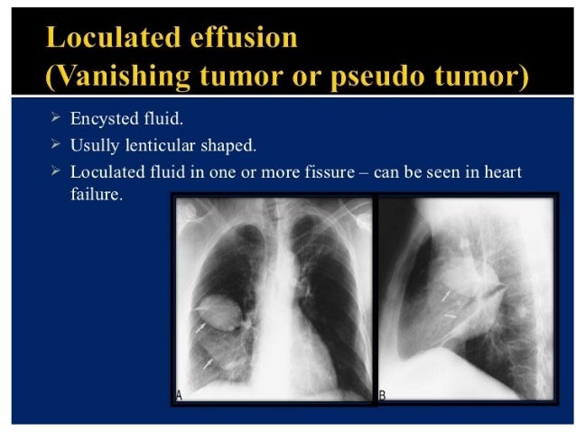
Pleural diseases chest radiology part1 from image.slidesharecdn.com Differentiate from an elevated hemidiaphragm. Parapneumonic effusion is defined as fluid in the pleural space in the presence of pneumonia, lung abscess, or bronchiectasis. In thoracic empyema (te) and complicated parapneumonic effusions. Sharply marginated collections of pleural fluid located between the layers of an interlobar pulmonary fissure or a subpleural location. It is just loculated pleural effusion that causes shadowing of the left middle and lower lung field. Pleural effusions are abnormal accumulations of fluid within the pleural space. Case contributed by dr prashant mudgal. Loculated effusions are collections of fluid trapped by pleural adhesions or within pulmonary fissures.
Parapneumonic effusion is defined as fluid in the pleural space in the presence of pneumonia, lung abscess, or bronchiectasis.
Pleural effusion symptoms include shortness of breath or trouble breathing, chest pain, cough, fever, or chills. For the radiographer there can be more to imaging a pleural effision than you might think. The opacity is effusion is sometimes hard to smoothly marginated and biconvex. A rational diagnostic workup, emphasizing the most common causes. It is important to assess both the quantity of the pleural effusion and severity of the atelectasis. Treatment of loculated pleural effusion with intrapleural urokinase in children. Consult surgery or interventional radiology for bleeding from tumors or vascular pathology. Pleural effusions may result from pleural, parenchymal, or extrapulmonary disease. Under normal conditions, pleural fluid is secreted by the parietal pleural capillaries at a rate of 0.01 millilitre per kilogram weight per hour. As the subpulmonic effusion grows in size, it first fills and thus blunts the posterior costophrenic sulcus, visible on the lateral chest. Pleural effusions are classified as either. There is blunting of both costophrenic angles, right greater than left. Diffuse nodules and opacification in right lung with compressive.
Pleural effusions demonstrated with chest radiography are nothing if not commonplace loculated pleural effusion. [diagnosis of pleural effusions and atelectases:


0 Comments